
You have no items in your shopping cart.
Protocol Optimization & Best Practices
9 Tips for Successful Immunohistochemistry
Practical strategies to optimize antigen retrieval, block non-specific background, and choose detection systems.
Immunohistochemistry (IHC) is an essential technique in clinical diagnostics and biological research. By using labeled antibodies to target specific tissue antigens in situ, IHC enables clear visual localization and spatial characterization of proteins within intact tissue architecture.
Combined with optimized chromogenic or fluorescent staining, IHC provides quantitative data regarding biomarker distribution, tissue orientation, cellular expression levels, and disease pathology. Protocol parameters heavily influence staining quality—careful optimization saves valuable time, prevents sample loss, and ensures publication-ready results.
Comprehensive Protocol Sheet
Need exact incubation times, buffer formulas, or step-by-step bench instructions? Access Biorbyt's official Immunohistochemistry Protocol Guide.
1. Applications of Immunohistochemistry
IHC is utilized worldwide across clinical pathology, oncology, translational medicine, and drug development:
- Pathology & Cancer Diagnostics: Identifying cellular origins, grading tumor margins, and classifying specific subtypes (e.g., HER2, ER/PR, PD-L1).
- Drug Discovery & Biomarker Profiling: Evaluating target engagement, measuring tissue distribution of biotherapeutics, and monitoring pharmacodynamic responses.
- Multiparametric Tissue Analysis: Visualizing spatial relationships between distinct cell populations (e.g., tumor-infiltrating lymphocytes vs. stroma).
2. Basic Workflow Overview
A standard IHC experiment follows three fundamental stages:
- 1. Tissue Preparation: Formalin-fixing and embedding in paraffin (FFPE / IHC-P) or flash-freezing in OCT compound (Frozen / IHC-Fr).
- 2. Immunostaining: Antigen retrieval, blocking non-specific sites, and primary antibody incubation targeting the protein of interest.
- 3. Detection & Visualization: Signal development using enzyme-conjugated secondary systems (HRP/DAB or AP/Fast Red) or fluorescent dyes, followed by microscopic examination.
3. Nine Tips to Successful IHC
Select & Test Validated Antibodies
Verify whether an antibody is validated for paraffin sections (IHC-P) or frozen tissue (IHC-Fr). Review species cross-reactivity datasheets and test new primary antibodies across a dilution range to establish optimal stain intensity with minimal background. Utilize matching diluent buffers to maintain antibody stability.
Match Tissue Preparation to Method
Choose between FFPE and frozen sections based on your target antigen: FFPE preserves tissue morphology best but can mask epitopes, whereas cryosections preserve native protein conformation better but exhibit poorer morphological detail. Use a microtome for uniform FFPE sectioning and a cryostat for frozen samples.
Incorporate Robust Positive & Negative Controls
Always include known positive tissue controls (expressing target antigen) and negative controls (omitting primary antibody or using target-knockout tissue) in every run. Controls confirm staining specificity and simplify troubleshooting when unexpected results arise.
Optimize Fixation Conditions
Fixative type, concentration, pH, and exposure duration strongly influence epitope preservation. Over-fixation creates excessive protein cross-linking that masks epitopes, whereas under-fixation causes tissue autolysis and uneven edge staining.
Perform Appropriate Antigen Retrieval (HIER / PIER)
Formalin fixation masks epitopes. Unmask targets using Heat-Induced Epitope Retrieval (HIER) in acidic citrate buffer (pH 6.0) or alkaline Tris-EDTA buffer (pH 9.0), or Enzymatic Retrieval (PIER) using proteinase K or trypsin. Test both pH ranges to determine optimal retrieval efficiency.
Block Endogenous Enzymatic Activity
Inactivate endogenous tissue enzymes prior to detection. Quench endogenous peroxidases using 3% hydrogen peroxide (H2O2) in methanol or water when using HRP detection systems. Block endogenous biotin or alkaline phosphatase when using avidin-biotin or AP detection complexes.
Prevent Non-Specific Fc Binding
Block non-specific antibody binding to cell surface Fc receptors by incubating sections with 5%–10% normal serum obtained from the host species of the secondary antibody, or apply a specialized background blocking reagent prior to primary antibody incubation.
Choose the Right Detection System
Select direct detection (fluorophore-conjugated primary) for highly abundant targets or multiplexing. Select indirect detection (unconjugated primary + polymer/HRP-secondary conjugate) for low-abundance targets to amplify signal strength. Polymer-based HRP systems eliminate endogenous biotin interference entirely.
Optimize Chromogen & Counterstain Pairing
Match your chromogen substrate (e.g., DAB brown, AEC red) with an appropriate counterstain (e.g., Hematoxylin) to provide high visual contrast. Ensure mounting media is chemically compatible with your chosen chromogen (organic solvent-based vs. aqueous mounting media).
4. Validated IHC Staining Gallery
Examples of specific, low-background immunohistochemical staining using Biorbyt primary antibodies:
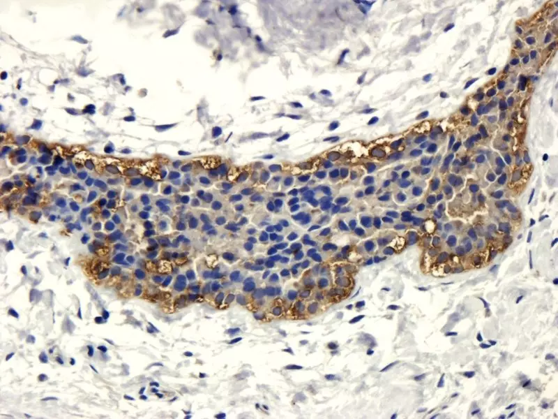
Human mammary fibroma tissue stained with anti-CD206 / CD133 (orb99113, primary dilution 1:100).
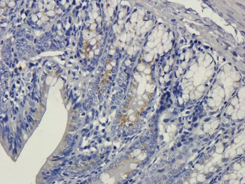
IHC-P staining of rat colon tissue using anti-VEGFA antibody (orb256347, 5 μg/ml).
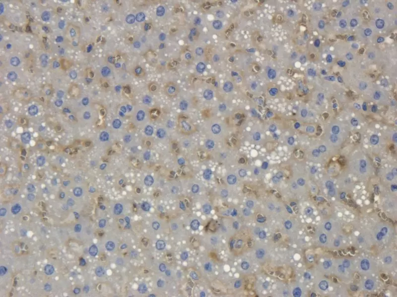
IHC-P staining of rat liver tissue using anti-Collagen X antibody (orb10444, 2.5 μg/ml).
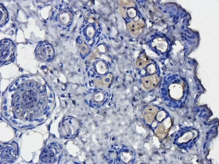
IHC-P staining of paraffin-embedded mouse skin tissue using anti-CD163 antibody (orb13303, primary dilution 1:200).
Need Assistance Optimizing Your Protocol?
Biorbyt’s scientific support team offers protocol optimization guidance and positive control recommendations tailored to your research target.
Contact Technical Support